
|
Microscope - Probe Display > Dr. Probe / Documentation / GUI / Microscope |
|
Parameters of the probe function display are set using the display section.
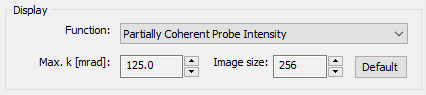
Select the active probe function to be displayed in the attached window from the dropdown list. Next to a diversity of probe functions, you can also display the aberration function and a simulated ronchigram of an artificial thin amorphous object. Each function is calculated using the current set of microscope parameters as well as with the two imaging parameters Max. k and Image size specified in the display section.
The physical sampling of the probe function is set by specifying the maximum diffraction vector k in units of milliradians. The current setting for this parameter is displayed and the number can be adjusted using the attached spin button controls. By increasing the max. diffraction vector, the image is zoomed to a finer scale for probe functions shown in real space, and to a coarser scale for functions shown in Fourier-space.
The number of image pixels used to sample the probe function in the display can be changed also with the attached spin controls.
Press the [Default] button to reset the max. diffraction vector and the image size to reasonable values for the currently selected probe function.
Last update: Jan 27, 2019 contact disclaimer(de)